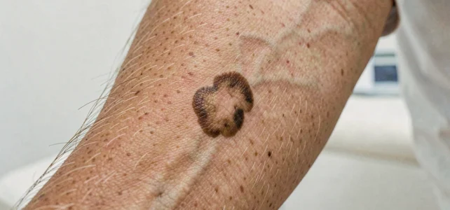
Skin cancer encompasses several types of skin tumors, the visual aspects of which vary considerably. Basal cell carcinoma, squamous cell carcinoma, melanoma: each presents distinct signs on the skin, and their early recognition directly influences the prognosis. Comparing a suspicious lesion to visual references remains a common reflex, but this approach has limitations that must be understood.
Limitations of online photos for identifying skin cancer
Images of skin cancers available on the web often show lesions at an advanced stage, on light skin. This visual bias poses a concrete problem: an early-stage melanoma does not resemble the most widely circulated photos. On medium or dark skin, a basal cell carcinoma may appear as a common pigmented spot rather than a translucent pearl.
Image galleries typically classify cancers by type (basal cell, squamous cell, melanoma), which gives the impression of a clear distinction. In practice, benign lesions such as seborrheic keratosis or an angioma can mimic the appearance of cancer, and vice versa. Relying solely on a visual comparison with photos and images of skin cancer does not replace a dermoscopic examination conducted by a specialist.
Mobile applications for analyzing skin lesions using artificial intelligence illustrate this difficulty. According to a recent analysis, these tools exhibit variable performance depending on phototypes and shooting conditions. An expert dermatologist remains necessary to validate or refute any suspicion, even when the algorithm assesses a lesion as “low risk.”
Warning signs of melanoma: beyond the ABCDE rule
The ABCDE rule (Asymmetry, Border irregularity, Color heterogeneity, Diameter, Evolution) is the foundation of self-monitoring moles. It appears in almost all public guides, rightly so. However, it does not cover all scenarios.
The most discriminating criterion remains the rapid evolution of a lesion over a few weeks or months. A mole that changes shape, color, bleeds spontaneously, or itches warrants medical advice, even if it does not meet the other ABCDE criteria.
One lesser-known sign deserves attention:
- The “ugly duckling”: a nevus that distinctly differs from all other moles in the same body area, by its size, color, or texture, justifies examination even if it appears regular when viewed in isolation.
- Achromic melanomas (without dark pigmentation): they appear pinkish or reddish and often escape classic visual detection, particularly on very light skin.
- Unusual locations: scalp, soles of the feet, under the nails, mucous membranes. These areas are rarely self-examined, and cancers that develop there are often diagnosed later.
Basal cell carcinoma and squamous cell carcinoma: distinct signals from melanoma
Basal cell carcinoma accounts for the majority of skin cancers. It typically manifests as a small, pearly, translucent lesion, sometimes featuring fine telangiectasias (visible blood vessels). It can also appear as a wound that has not healed for several weeks, or as a crusty area that bleeds and then reforms.
Squamous cell carcinoma, less common, often appears on sun-exposed areas: face, ears, backs of hands. It presents as a firm nodule, sometimes ulcerated, or as a thickened red plaque. Its particularity: it can develop on a pre-existing actinic keratosis, those small rough and dry spots that are sometimes felt more than seen.
Actinic keratosis is not cancer, but a precancer. Its presence indicates cumulative sun exposure and an increased risk of transformation. Regular monitoring is an integral part of skin screening.
Teledermatology and screening pathways: what is changing
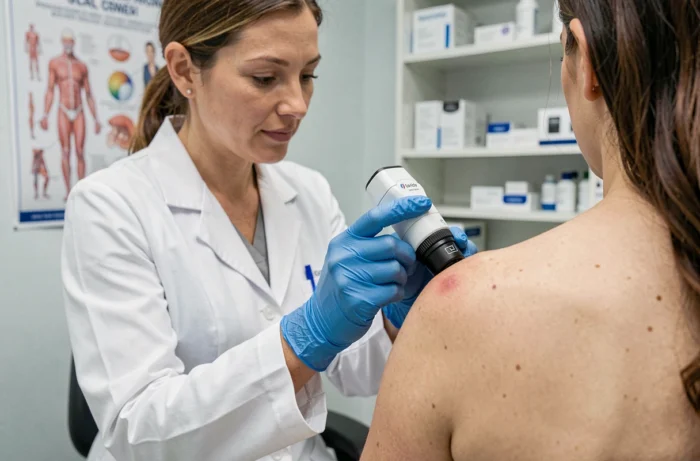
Access to a dermatologist remains unequal across regions. Teledermatology is gradually changing this pathway. The principle: the primary care physician photographs a suspicious lesion (with or without a portable dermoscope) and sends the images to a dermatologist for remote advice.
Recent studies indicate that this circuit shortens the time between suspicion and specialized consultation. In some trials, melanomas detected via teledermatology had a lower Breslow thickness, indicating a diagnosis at an earlier stage. The available data do not yet allow for a conclusion about a generalizable benefit, but the trend is consistent with the goals of rapid detection.
Meanwhile, discussions are ongoing in Europe to adapt the frequency of screening to individual risk levels. Germany, for example, plans to revise its skin screening program by 2027 to move to a risk-adapted screening model. Individuals with light phototypes, family histories, or a high number of nevi would be examined more frequently, while low-risk profiles could space out their check-ups.
Self-monitoring of the skin: a concrete method
Regularly photographing moles with a smartphone allows for documenting their evolution over time. This practice, sometimes referred to as “home mole mapping,” facilitates comparison from month to month and provides the dermatologist with a usable visual history during the consultation.
Some guidelines to structure this self-monitoring:
- Examine the entire body every three months, including the back (using a mirror or a friend), the scalp, and the spaces between the toes.
- Photograph each suspicious lesion with a graduated ruler next to it to maintain a size reference.
- Note the date of each photo and systematically compare it with the previous image to spot any changes in color, border, or relief.
The change in appearance remains the most reliable warning signal, regardless of the type of suspected skin cancer. A lesion that has been stable for years is more reassuring than a spot that has appeared recently, even if it seems regular. In case of doubt, consultation remains the only way to obtain a reliable diagnosis through dermoscopy or biopsy.